Retinal Tear Optos : Retinal Holes And Tears Recognizing Pathology Optos / A scan to show a healthy eye or detect disease.. Uveitis is inflammation of the uveal tract which can be caused by autoimmune disorders such as rheumatoid arthritis or infection which can be idiopathic. It shows the retina (where light and images hit), the optic disk (a spot on the retina that holds the optic nerve, which sends. Another condition leading to retinal detachment is a giant retinal tear, defined as a retinal tear that extends 3 clock hours (90 degrees) or more around the circumference of the globe. But, diseases such as macular degeneration, glaucoma, retinal tears or detachments, and other health problems such as diabetes and high blood pressure can be seen with a thorough exam of the retina. The optomap® retinal exam produces an image that is as unique as you fingerprint and provides us with a wide view to look at the health of your retina.
Tearing of the retina may produce retinal or vitreous hemorrhages. A scan to show a healthy eye or detect disease. Operculated retinal tear an operculated tear results from vitreous traction that pulls a plug of sensory retina out into the vitreous cavity (see illustration). +61 8 8444 6500 auinfo@optos.com building the retina company optos.com opto map color image highlighting both the retinal tear and pvd, both in the far periphery. Retinal tears when a retinal tear or hole hasn't yet progressed to detachment, your eye surgeon may suggest one of the following procedures to prevent retinal detachment and preserve vision.
Widefield And Ultra Widefield Imaging When And Why To Use Them from s3-ap-southeast-2.amazonaws.com The most common reason for a rd is a retinal tear and the most common reason for a tear is a pvd. Operculated retinal tear an operculated tear results from vitreous traction that pulls a plug of sensory retina out into the vitreous cavity (see illustration). +1 508 787 1400 usinfo@optos.com optos australia tel: Retinal tears when a retinal tear or hole hasn't yet progressed to detachment, your eye surgeon may suggest one of the following procedures to prevent retinal detachment and preserve vision. There are many varieties of tears and breaks, but we can see them all with the optos and prescribe proper treatment accordingly. But, diseases such as macular degeneration, glaucoma, retinal tears or detachments, and other health problems such as diabetes and high blood pressure can be seen with a thorough exam of the retina. Some times the retinal vessels on the detachment will be dark in appearance, as seen in a retinoschisis. These types of retinal detachments are the most common.
+61 8 8444 6500 auinfo@optos.com building the retina company optos.com opto map color image highlighting both the retinal tear and pvd, both in the far periphery.
Some times the retinal vessels on the detachment will be dark in appearance, as seen in a retinoschisis. A giant retinal tear typically develops slightly posterior and parallel to the ora serrata, with the vitreous base attached to the anterior margin of the tear. We offer the optomap® retinal exam as an important part of our temple eye exams. An optomap® retinal exam provides: A retinal detachment (rd) is the separation of the sensory retina from the retinal pigment epithelium (rpe) (outer segments of the photoreceptors from the microvilli of the rpe). An optomap® retinal exam provides: The most frequent cause of an operculated tear is a pvd. Another condition leading to retinal detachment is a giant retinal tear, defined as a retinal tear that extends 3 clock hours (90 degrees) or more around the circumference of the globe. +61 8 8444 6500 auinfo@optos.com building the retina company optos.com opto map color image highlighting both the retinal tear and pvd, both in the far periphery. The optomap® retinal exam produces an image that is as unique as you fingerprint and provides us with a wide view to look at the health of your retina. But, diseases such as macular degeneration, glaucoma, retinal tears or detachments, and other health problems such as diabetes and high blood pressure can be seen with a thorough exam of the retina. Optos retinal imaging the optos imaging technology allows us to give our patients a more thorough health evaluation. The retina first tears, and then it detaches.
But, diseases such as macular degeneration, glaucoma, retinal tears or detachments, and other health problems such as diabetes and high blood pressure can be seen with a thorough exam of the retina. Optos retinal imaging the optos imaging technology allows us to give our patients a more thorough health evaluation. It can occur at any age and can result from many causes including systemic disease. Retinal defects come in different shapes and sizes and may be either partial or full thickness. But, diseases such as macular degeneration, glaucoma, retinal tears or detachments, and other health problems such as diabetes and high blood pressure can be seen with a thorough exam of the retina.
Safety And Efficacy Of The Use Of Navigated Retinal Laser As A Method Of Laser Retinopexy In The Treatment Of Symptomatic Retinal Tears Eye from media.springernature.com Tearing of the retina may produce retinal or vitreous hemorrhages. These types of retinal detachments are the most common. Optos retinal photography many eye problems can develop without you knowing, in fact, you may not even notice any change in your sight, diseases or damage such as macular degeneration, glaucoma, retinal tears or detachments, and other health problems such as diabetes and high blood pressure can be seen with a thorough exam of the retina. Our astoria optometrists are able to monitor freckles in the back of the eye known as a choroidal nevus. Operculated retinal tear an operculated tear results from vitreous traction that pulls a plug of sensory retina out into the vitreous cavity (see illustration). Almost all clinically significant rds (greater than 2 disc diameters (dd) from the edge of the break or 1 dd posterior to the equator) if left untreated will eventually lead to a. The most common reason for a rd is a retinal tear and the most common reason for a tear is a pvd. The optomap® retinal exam produces an image that is as unique as you fingerprint and provides us with a wide view to look at the health of your retina.
Optos retinal photography many eye problems can develop without you knowing, in fact, you may not even notice any change in your sight, diseases or damage such as macular degeneration, glaucoma, retinal tears or detachments, and other health problems such as diabetes and high blood pressure can be seen with a thorough exam of the retina.
The optomap® retinal exam produces an image that is as unique as you fingerprint and provides us with a wide view to look at the health of your retina. +1 508 787 1400 usinfo@optos.com optos australia tel: We offer the optomap® retinal exam as an important part of our temple eye exams. A scan to show a healthy eye or detect disease. Another condition leading to retinal detachment is a giant retinal tear, defined as a retinal tear that extends 3 clock hours (90 degrees) or more around the circumference of the globe. It is important to detect problems in the eye, like diabetes, macular degeneration, melanoma, bleeding, hypertensive retinopathy and glaucoma before they cause loss of vision. These types of retinal detachments are the most common. A giant retinal tear typically develops slightly posterior and parallel to the ora serrata, with the vitreous base attached to the anterior margin of the tear. If you think that you are suffering from a retinal tear, please call your optometrists at the vision center in fort worth and lubbock, tx. Operculated retinal tear an operculated tear results from vitreous traction that pulls a plug of sensory retina out into the vitreous cavity (see illustration). Retinal imaging takes a digital picture of the back of your eye. A scan to show a healthy eye or detect disease. Uveitis is inflammation of the uveal tract which can be caused by autoimmune disorders such as rheumatoid arthritis or infection which can be idiopathic.
But, diseases such as macular degeneration, glaucoma, retinal tears or detachments, and other health problems such as diabetes and high blood pressure can be seen with a thorough exam of the retina. These types of retinal detachments are the most common. Tearing of the retina may produce retinal or vitreous hemorrhages. It shows the retina (where light and images hit), the optic disk (a spot on the retina that holds the optic nerve, which sends. The optomap® retinal exam produces an image that is as unique as you fingerprint and provides us with a wide view to look at the health of your retina.
Retinal Detachment Recognizing Pathology Optos from recognizingpathology.optos.com The optomap® retinal exam produces an image that is as unique as you fingerprint and provides us with a wide view to look at the health of your retina. The device allows for the discovery of retinal melanomas and. Retinal tears when a retinal tear or hole hasn't yet progressed to detachment, your eye surgeon may suggest one of the following procedures to prevent retinal detachment and preserve vision. The most common reason for a rd is a retinal tear and the most common reason for a tear is a pvd. Optos retinal imaging the optos imaging technology allows us to give our patients a more thorough health evaluation. But, diseases such as macular degeneration, glaucoma, retinal tears or detachments, and other health problems such as diabetes and high blood pressure can be seen with a thorough exam of the retina. Operculated retinal tear an operculated tear results from vitreous traction that pulls a plug of sensory retina out into the vitreous cavity (see illustration). This combined device facilitates the early detection, management and effective treatment of disorders and diseases evidenced in the retina such as retinal detachments and tears, glaucoma, diabetic retinopathy.
Optos retinal imaging the optos imaging technology allows us to give our patients a more thorough health evaluation.
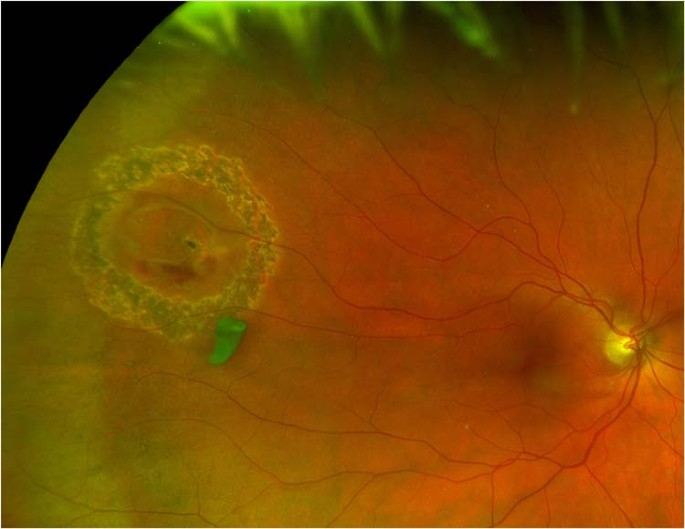
Retinal tear temporal about 4 o'clock in the left eye. Operculated retinal tear an operculated tear results from vitreous traction that pulls a plug of sensory retina out into the vitreous cavity (see illustration). An optomap® retinal exam provides: +61 8 8444 6500 auinfo@optos.com building the retina company optos.com opto map color image highlighting both the retinal tear and pvd, both in the far periphery. Operculated retinal tear an operculated tear results from vitreous traction that pulls a plug of sensory retina out into the vitreous cavity (see illustration). A scan to show a healthy eye or detect disease. The optomap® retinal exam produces an image that is as unique as you fingerprint and provides us with a wide view to look at the health of your retina. If you think that you are suffering from a retinal tear, please call your optometrists at the vision center in fort worth and lubbock, tx. Post laser treatment is present. The optos daytona helps our doctors diagnose eye diseases such as glaucoma, macular degeneration, and diabetic retinopathy. Almost all clinically significant rds (greater than 2 disc diameters (dd) from the edge of the break or 1 dd posterior to the equator) if left untreated will eventually lead to a. She immediately followed up with a retinal specialist who performed an emergency laser surgery that successfully repaired the tear. The uvea is responsible for the transport of blood including inflammatory cells and is critical for vision.
A retinal tear, or retinal detachment, occurs when the retina is torn from the underlying tissue retinal tear. Retinal holes and tears are commonly encountered during dilated fundus examination of both symptomatic and asymptomatic patients.


0 Komentar